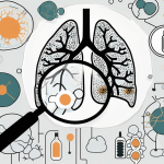
Diagnosing lung cancer accurately is paramount, and tissue biopsy stands as a critical tool in this process. This article breaks down the biopsy’s vital role, detailing various procedures like bronchoscopy and needle biopsy, complemented by imaging techniques like CT scans for precision. It outlines the biopsy process from patient preparation through to the analysis of samples by pathologists. While acknowledging the risks, the article emphasizes biopsy’s unparalleled value in confirming cancer presence, type, and stage, guiding tailored treatment plans. It aims to demystify the process, highlighting advancements in biopsy techniques and addressing patient concerns, ultimately underscoring the procedure’s significance in effective lung cancer management.
Suspected lung cancer can be a distressing and overwhelming experience. When it comes to diagnosing this potentially life-threatening condition, a tissue biopsy plays a crucial role. In this article, we will explore the importance of tissue biopsy in diagnosing lung cancer, the different types of biopsy procedures available, the role of imaging techniques in guiding the biopsy, and the step-by-step process of conducting a tissue biopsy. We will also delve into the risks and complications associated with the procedure, the accuracy and reliability of tissue biopsy in detecting lung cancer, advancements in biopsy techniques, the role of pathologists in analyzing biopsy samples, and patient experiences and expectations during the procedure. By the end, you will have a better understanding of how this diagnostic tool can aid in the detection and treatment of suspected lung cancer.
Understanding the Importance of Tissue Biopsy in Diagnosing Lung Cancer
In order to accurately diagnose suspected lung cancer, doctors often need to analyze samples of lung tissue. This is where tissue biopsy becomes invaluable. A tissue biopsy involves the surgical removal of a small piece of lung tissue for examination under a microscope. By analyzing the cells in the sample, pathologists can determine if cancer is present and, if so, the type and stage of the disease.
When a patient presents with symptoms that may indicate lung cancer, such as persistent cough, shortness of breath, or unexplained weight loss, a tissue biopsy is often recommended. The procedure is typically performed by a thoracic surgeon or an interventional radiologist, who will use imaging techniques like CT scans or ultrasound to guide the biopsy needle to the precise location of the suspicious tissue. This ensures that the sample obtained is representative of the area of concern.
Once the tissue sample is obtained, it is sent to a pathology laboratory for analysis. Pathologists, who are specialized doctors trained in diagnosing diseases by examining cells and tissues, carefully study the sample under a microscope. They look for abnormal cell growth, changes in cell structure, and other features that may indicate the presence of cancer.
It is important to note that a tissue biopsy is the gold standard for diagnosing lung cancer. While imaging techniques like CT scans and X-rays can provide valuable information, only a tissue biopsy can provide a definitive diagnosis. By confirming the presence of cancer and identifying its specific characteristics, doctors can tailor treatment plans to individual patients, leading to more effective outcomes.
Furthermore, a tissue biopsy can also provide additional information beyond just the presence of cancer. Pathologists can determine the type of lung cancer, such as non-small cell lung cancer or small cell lung cancer, which is crucial for selecting the most appropriate treatment approach. Additionally, the biopsy can reveal the stage of the disease, indicating how far the cancer has spread within the lungs or to other parts of the body. This information helps doctors determine the prognosis and develop a personalized treatment plan.
While a tissue biopsy is generally a safe procedure, there are some risks involved. These may include bleeding, infection, or damage to surrounding structures. However, the benefits of obtaining an accurate diagnosis far outweigh the potential risks. By identifying lung cancer early through a tissue biopsy, patients have a better chance of receiving timely and effective treatment, improving their overall prognosis.
In conclusion, tissue biopsy plays a crucial role in diagnosing lung cancer. It provides a definitive diagnosis, identifies the type and stage of the disease, and guides treatment decisions. By understanding the importance of tissue biopsy, patients and healthcare providers can work together to ensure the best possible outcomes for individuals facing a suspected lung cancer diagnosis.
Different Types of Tissue Biopsy Procedures
When it comes to diagnosing and treating various medical conditions, tissue biopsy procedures play a crucial role. These procedures allow doctors to obtain samples of abnormal tissue for further analysis, helping to determine the presence of diseases such as cancer. However, not all tissue biopsy procedures are the same, as each one has its own advantages and considerations.
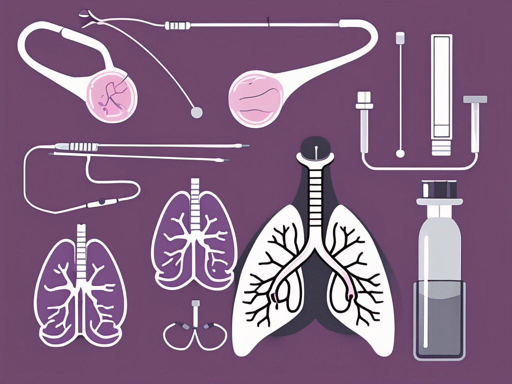
One commonly used method is the bronchoscopy biopsy. This procedure involves the insertion of a thin tube called a bronchoscope through the nose or mouth and into the lungs. The bronchoscope allows the doctor to visually examine the airways and collect samples from any abnormal-looking areas. This type of biopsy is particularly useful for diagnosing lung conditions and detecting tumors in the respiratory system.
Another option is the needle biopsy, which can be done either through the skin or with the assistance of imaging techniques such as CT or ultrasound. During a needle biopsy, a thin needle is used to extract tissue samples from the lung. This procedure is less invasive compared to surgical biopsies and can be performed on an outpatient basis. It is commonly used to diagnose lung nodules, which are small growths in the lung tissue that may be cancerous or non-cancerous.
However, in cases where other biopsy procedures have not yielded sufficient samples or when a larger tissue sample is needed for further analysis, surgical biopsy may be necessary. This procedure involves creating a small incision in the chest and using specialized instruments to remove tissue from the lung. Surgical biopsies are typically performed in a hospital setting under general anesthesia. They allow for a more extensive examination of the tissue and are often used when there is a high suspicion of cancer or when the exact nature of the abnormality is unclear.
It is important to note that the choice of biopsy procedure depends on various factors, including the location and size of the suspected tumor, as well as the overall health of the patient. The medical team will carefully evaluate each case to determine the most appropriate biopsy method to ensure accurate diagnosis and effective treatment.
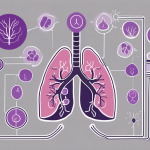
The Role of Imaging Techniques in Guiding Tissue Biopsy Procedures
Imaging techniques, such as CT scans and ultrasound, play a crucial role in guiding tissue biopsy procedures. By providing detailed images of the lungs and surrounding structures, these tools help doctors locate suspicious areas and guide the biopsy needle or bronchoscope to the precise location.
CT-guided needle biopsy is particularly useful when the suspected tumor is in a hard-to-reach area or located deep within the lung. This procedure combines real-time imaging with needle biopsy, allowing doctors to accurately target the abnormal tissue. Ultrasound-guided biopsies, on the other hand, use sound waves to create images of the lung and guide the needle to the desired location. The use of imaging techniques during biopsy procedures not only increases accuracy but also minimizes the risk of complications.
Step-by-Step Guide to Conducting a Tissue Biopsy for Suspected Lung Cancer
The process of conducting a tissue biopsy for suspected lung cancer typically involves several key steps:
- Patient preparation : Before the procedure, your doctor will provide specific instructions to ensure you are ready and comfortable. This may include fasting for a certain period, avoiding certain medications, and discussing any allergies or medical conditions.
- Anesthesia : Depending on the type of biopsy, your doctor may administer local anesthesia to numb the area or general anesthesia to ensure you are asleep and pain-free during the procedure.
- Biopsy procedure : The chosen biopsy method will be performed, whether it is a bronchoscopy, needle biopsy, or surgical biopsy. The doctor will carefully extract a sample of tissue from the suspected area for further analysis.
- Recovery and observation : After the biopsy, you will be taken to a recovery area, where you will be monitored for any immediate complications or side effects from the procedure.
- Analysis and diagnosis : The tissue sample will be sent to a pathology lab, where a pathologist will analyze the cells, looking for any abnormalities or signs of cancer. This process may take several days, and the results will be shared with you by your doctor.
- Treatment planning : Once a definitive diagnosis has been made, your doctor will discuss the treatment options available to you and develop a personalized plan based on your specific condition.